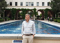


Dr Andres Collazo
Director of the Biological Imaging Facility The overarching goal of my research program is to push the limits of optical microscopy to study how proteins function in as close to in vivo conditions as possible. A fascinating area of biology is the study of single protein molecule dynamics in living animals, a potentially powerful means of identifying abnormal protein function and understanding molecular causes of medical disorders. Fluorescence fluctuation spectroscopy (FFS) encompasses several techniques, including fluorescence correlation spectroscopy (FCS) and photon counting histograms (PCH), which provide quantitative data on the dynamics of single protein molecules in living cells (SLAUGHTER et al. 2007; SLAUGHTER AND LI 2010). These techniques detect fluorescence intensity fluctuations as labeled molecules pass in and out of a small confocal volume (<1 femtoliter), created by focusing a low intensity laser. FCS can determine the mobility and concentration of fluorescently labeled proteins (HAUSTEIN AND SCHWILLE 2008; RIES et al. 2009; SCHWILLE AND RIES 2011). Only rarely have these techniques been applied in vivo to multicellular animals (RIES et al. 2009; YU et al. 2009). We have used FCS in living zebrafish embryos to show that changes in α-catenin mobility during development of the pharyngeal pouch reflects the adherens junction remodeling underlying pouch formation (CHOE et al. 2013). Bachelors of Science in Biology from Cornell University, Ph.D in Zoology from the University of California at Berkeley.
Contact Info Office: 626-395-2761
BIF: 626-395-2863
acollazo at caltech.edu
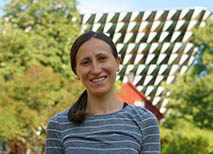
Dr Zhongying (Ying) Wang
Microscopy Application Scientist I completed my graduate training in Molecular Biology at the University of Southern California (USC), where I focused on live-cell imaging of pancreatic beta cell metabolism using fluorescence lifetime imaging microscopy (FLIM). My research centered on using endogenous autofluorescence to monitor real-time metabolic changes in living cells. This work sparked a deep interest in applying advanced imaging techniques to understand cellular function.
During my postdoctoral training at UCLA and later at USC, I continued studying diabetes development in mouse models. I developed in vivo imaging techniques using the anterior chamber of the eye, enabling non-invasive, longitudinal monitoring of pancreatic islets in live mice. In parallel, I expanded my expertise in both FLIM and STED microscopy. I’m excited to bring this imaging experience to Caltech and explore collaborative opportunities across disciplines. With a strong foundation in both imaging technology and mice research, I aim to support projects that can benefit from live, high-resolution visualization of biological processes in real time.
Contact Info BIF: 626-395-2863
zwang10 at caltech.edu